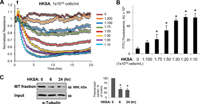
Figure 1.

HKSA-induced EC permeability is accompanied by MT destabilization. A, HPAEC monolayers grown on gold microelectrodes were challenged with the indicated concentrations of HKSA, and TER was monitored for 20 h. B, cells grown on 96-well plates with immobilized biotinylated gelatin were exposed to HKSA for 2 h. After completion of stimulation, FITC–avidin (25 μg/ml) was added to the cells, and they were incubated for 3 min followed by washing with PBS and measurement of FITC fluorescence in a Victor X5 plate reader. Normalized values are expressed as mean ± S.D.; n = 6, *, p < 0.05. C, MT fractionation from control and HKSA-treated (2 × 10⁸ particles/ml, 6 or 24 h) EC was performed by separation of soluble depolymerized tubulin and insoluble tubulin polymers assembled into MT by centrifugation as described under “Experimental procedures.” Densitometry results are shown as mean ± S.D. n = 5; *, p < 0.05.
