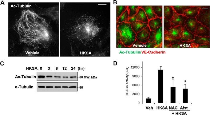
Figure 2.
HKSA decreases the pool of acetylated microtubules and increases HDAC activity in a redox-dependent manner. A, the levels of acetylated MT in control and HKSA-treated (2 × 10⁸ particles/ml, 6 h) cells were determined by immunofluorescence staining with acetylated tubulin antibody. Bar = 5 μm. B, co-staining of control and HKSA-stimulated (6 h) EC monolayers with antibodies to acetylated tubulin and VE-cadherin. VE-cadherin–positive adherens junctions outline the cell borders. Shown are representative results of three independent experiments. Bar = 10 μm. C, the total cell lysates collected from the cells were treated with HKSA for the indicated times, and acetylated tubulin levels in total cell lysates were analyzed by Western blotting (upper panel). Lower panel, shows total a-tubulin levels. Shown are representative results of three independent experiments. D, EC were stimulated with HKSA (3 h) followed by an HDAC6 activity assay. Where indicated, cells were pretreated for 30 min with the ROS scavenger N-acetylcysteine (NAC, 1 mm) or amifostine (Afst, amifostine trihydrate, WR2721, 4 mm). The results are presented after normalizing to background controls. *, p < 0.05; n = 5. Veh, vehicle.

