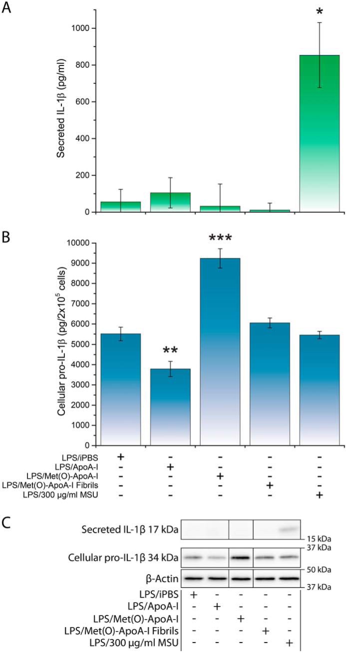
Figure 1.

Met(O)-ApoA-I did not activate cleavage of pro-IL-1β (34 kDa) and secretion of mature IL-1β (17 kDa) in murine macrophages. BMDMs were incubated with LPS (0.5 μg/ml) for 3.5 h (priming). The cell culture medium was then replaced with new medium containing LPS (0.5 μg/ml) and (as signal 2) apoA-I samples (1 μm) or monosodium urate crystals (MSU) (300 μg/ml) and incubated for an additional 4 h. MSU was used as a positive control for signal 2 induction (80). Results from a representative experiment (of three) are shown in the figure. Values are means and S.D. (error bars) of three independent determinations. t test significance values versus LPS/iPBS results are reported as p < 0.05 (*), p < 0.01 (**), and p < 0.001 (***). A, the concentration of mature IL-1β in incubation medium was measured by ELISA. B, pro-IL-1β levels were measured in cell extracts by Western blot analysis (see C) and are reported as pg of pro-IL-1β in cell extracts (from 2 × 10⁵ cells). C, representative Western blot analysis of cellular pro-IL-1β and secreted mature IL-1β with β-actin as a loading control. Vertical black lines in the Western blots indicate positions where unnecessary lanes were removed from the original scans.
