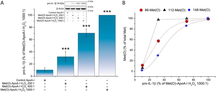
Figure 5.
Accumulation of pro-IL-1β (34 kDa) in BMDMs was proportional to the Met oxidation levels of Met(O)-ApoA-I. Different levels of Met oxidation were generated by oxidizing apoA-I with increasing concentrations of H2O2 (see Table 1). Unprimed BMDMs were incubated with apoA-I samples (1 μm) for 4 h. A, pro-IL-1β levels were measured in cell extracts by Western blotting and are expressed as a percentage of the pro-IL-1β levels induced by Met(O)-ApoA-I that was completely oxidized with a 1000-fold excess of H2O2 (absolute pro-IL-1β values obtained for this reference sample were 2086 ± 342 pmol of pro-IL-1β/2 × 105 cells (n = 4)). Results from a representative experiment (of two) are shown. Values are means and S.D. (error bars) of two independent determinations. t test significance values are reported as follows: ***, p < 0.001 for Met(O)-ApoA-I H2O2 200:1 versus iPBS, Met(O)-ApoA-I H2O2 500:1 versus Met(O)-ApoA-I H2O2 200:1, and Met(O)-ApoA-I H2O2 1000:1 versus Met(O)-ApoA-I H2O2 500:1. A representative Western blot analysis with β-actin as a loading control is shown in the inset. B, correlation between the extent of oxidation of each Met residue and the pro-IL-1β levels in the cell extracts.

