Figure 4.
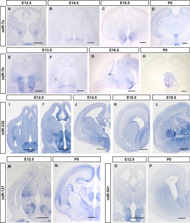
miRNAs are enriched in the developing mouse hypothalamus. In situ hybridization patterns for miR-7a (A–D), miR-7b (E–H), miR-335 (I–L), miR-137 (M and N), and miR-541 (O and P) in sections during brain development. A–D, the expression patterns of miR-7a in coronal sections of mouse brain at E12.5 (A), E14.5 (B), E16.5 (C), and E18.5 (D). E–H, miR-7b expression in coronal (E, F, and H) and sagittal sections (G) of mouse brain at E13.5, E16.5 and P0. I–L, O, and P, frontal sections through the telencephalon of E12.5-P0 mice, at a rostral or middle level of the pallidum, showing the dynamic expression of the miR-335 (I–L) and miR-541 (O and P) in the hypothalamus. The material shown corresponds to E12.5 (I, I′, and O), E14.5 (J), E16.5 (K and N), E18.5 (L), and P0 (P). M and N, in situ hybridization of miR-137 shows its expression pattern in the brain at E12.5 (M) and P0 (N). Scale bars in A–P are 500 μm.

