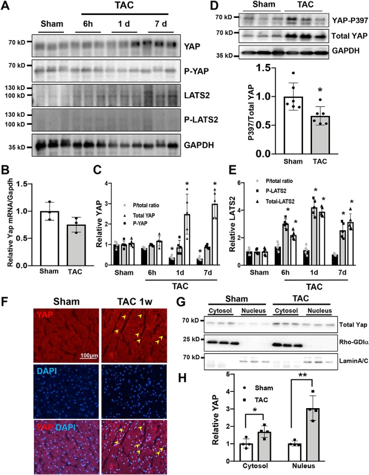
Figure 1.
YAP is activated after acute PO. Wildtype (WT) mice were subjected to TAC. The hearts were harvested at indicated time points (6 h to 7 days (d)). A, immunoblot analyses were performed to determine the phosphorylation status of YAP and Last2 in ventricular lysates. n = 5. B, mRNA expression of YAP in the mouse heart was measured by quantitative real-time PCR assays. n = 3. C, quantification of Ser-127–phosphorylated YAP and total YAP. D, immunoblot analyses of Ser-397–phosphorylated YAP and total YAP. E, quantification of phosphorylated Lats2 and total Lats2. F, immunostaining for YAP 7 days after TAC in the WT mouse heart. DAPI was used for nuclear staining. n = 4. G, subcellular localization of endogenous YAP in mouse ventricular extracts 7 days after TAC. Rho-GDIα and lamin A/C served as markers of cytosol- and nucleus-enriched fractions, respectively. n = 4. H, quantification of the data shown in G. The data are expressed as ratios relative to the mean value of the sham group. Data in graphs represent mean ± S.D.; *, p < 0.05; **, p < 0.01, compared with Sham. Statistical analyses were conducted with ANOVA or Student's t test. Post hoc analysis was conducted with Tukey's test.

