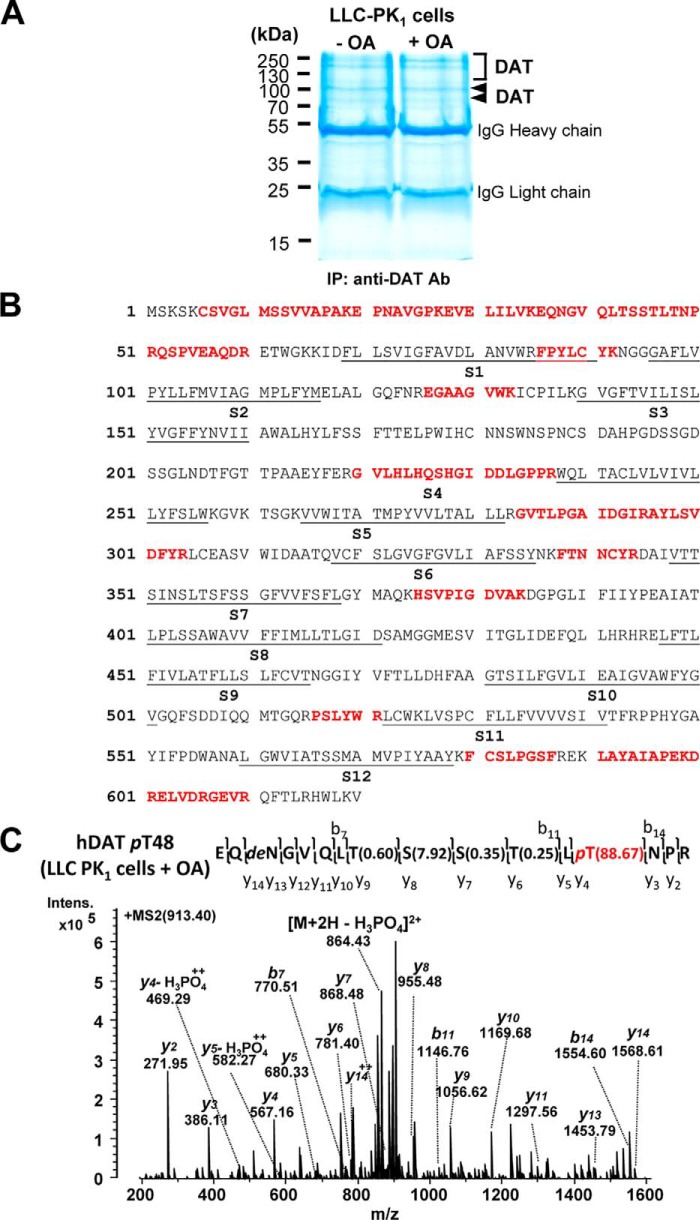
Figure 3.
Identification of PP1/2A-responsive DAT phosphorylation at Thr-48 by LC-MS/MS. A, representative colloidal blue–stained SDS-polyacrylamide gel image with proteins immunopurified by an anti-DAT antibody (Ab) from LLC-PK1 cells heterologously expressing hDAT in the absence (−) and presence (+) of OA. Arrowheads and square bracket, bands and region containing DAT protein identified by LC-MS/MS. The numbers refer to the positions of molecular weight markers. B, sequence coverage of human DAT with identified peptides (boldface type) by MS/MS from transfected LLC-PK1 cells. The putative transmembrane segments (S1–S12) of hDAT are underlined. C, MS/MS-identified hDAT phosphorylation site at Thr-48 (pT48) from transfected LLC-PK1 cells upon OA treatment. Shown is the MS/MS spectrum from the precursor ion at m/z 913.40 (2+) assigned to hDAT EQNGVQLTSSTLpTNPR (amino acids 36–51) with phosphorylation at Thr-48, which was verified by β-eliminated y-ions and neutral loss of H3PO4 from the precursor. The phosphosite localization probabilities determined by the Mascot delta score (38) are shown in parentheses. pT, phosphorylated threonine; deN, deamidated asparagine.

