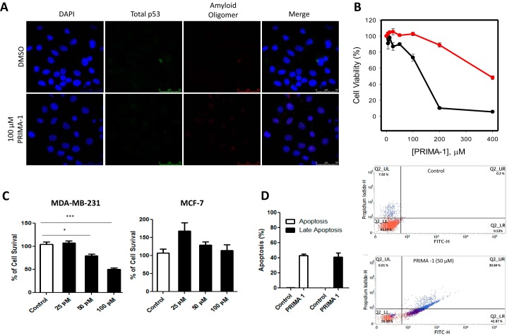
Figure 4.
A, MCF-7 cells were treated for 16 h with either 100 μm PRIMA-1 or 0. 1% DMSO as a control. The cells were labeled with anti-p53 (DO-1) and anti-oligomer (A11) antibodies, and nuclei were stained with DAPI. Columns from left to right show DAPI, p53 labeling, labeling of amyloid oligomers, and merged images. Magnification, ×400. Scale bar = 50 μm. B, MTT assay showing MDA-MB-231 (black line) and MCF-7 (red line) cell viability after treatment for 24 h with a serial dilution of PRIMA-1 from 0 (0.2% DMSO) to 200 μm. n = 4. C, cell proliferation assay using trypan blue showing a dose-dependent inhibition of cell proliferation after 24 h of treatment with PRIMA-1. n = 3 and significance marked with *(p < 0.05) and with *** (p < 0.001). D, AnnexinV/PI assay showing that PRIMA-1 induces apoptosis of MDA-MB-231 cells at 50 μm PRIMA-1, showing its effectiveness in this cell line in the concentration range used in this study.

