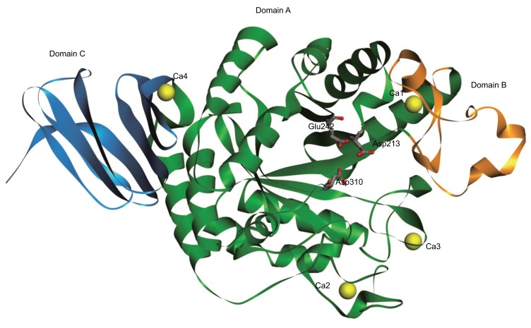
Figure 2.
Model structure of AGXA. The model structure of AGXA predicted by homology modeling based on the crystal structure of Anoxybacillus sp. SK3-4 α-amylase (PDB code: 5A2A). The three conserved active site residues are labeled by sticks. Domains A, B, and C are shown in green, orange, and blue, respectively. The calcium ions are shown in yellow. (The color version of the figure is available in the electronic copy of the article).

