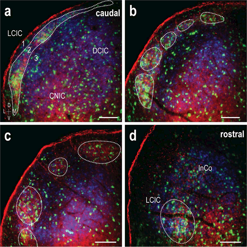
Figure 11.

Caudal to rostral overlap of GAD/EphA4/ephrin-B2 patterning. LCIC triple-labeling (GAD = green, EphA4 = red, ephrin-B2 = blue) throughout the caudorostral extent of the LCIC (a-d). Caudally, modules are interconnected (a), appearing as a strip along layer 2. In the mid-rostrocaudal zones the discontinuous modular morphology is most readily apparent (b, c), prior to converging and taking up a deeper position in the rostral extreme (d). Matching of GAD, EphA4, and ephrin B2 LCIC patterns is consistent throughout the caudorostral dimension. The observed shape changes are consistent with that previously described for other modular markers (Dillingham et al., 2017). Scale bars (a-d) = 100µm.
