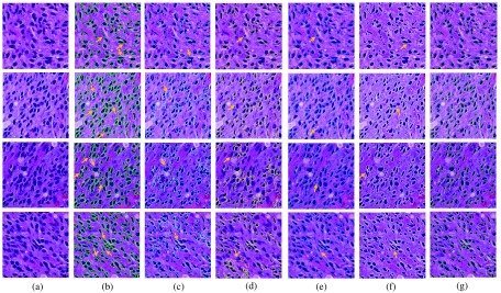
Fig. 8.
Comparison of segmentation results: (a) original image; (b) ground truth; (c) MS;38 (d) ISO;39 (e) SBGFRLS,40 (f) vanilla Chan–Vese model,1 and (g) our proposed method. We use arrows to demonstrate typical tissue areas, where our approach can correctly segment nuclei while other methods fail.

