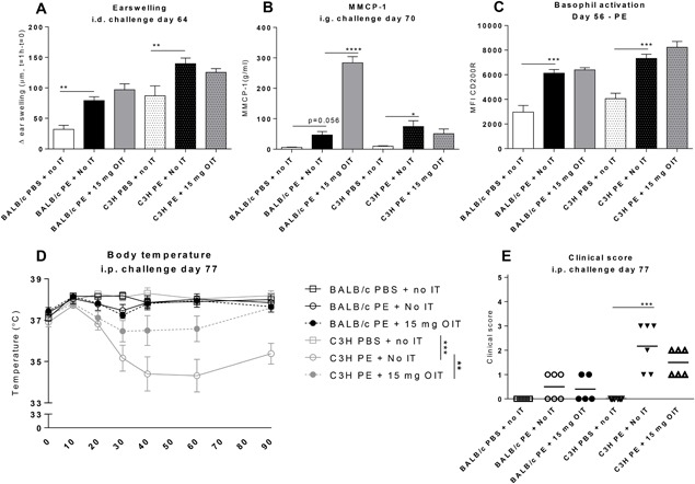
Figure 2.

Allergic manifestations in PE‐sensitized mice after receiving OIT. A, Acute allergic skin response measured as Δ ear swelling 1 h after i.d. challenge. B, Concentrations of MMCP‐1 in serum collected 30 min after i.g. challenge. C, On day 56 peripheral blood basophil activation was measured after whole blood stimulation with PE using flow cytometry. Cells were gated based on FSC‐SSC properties and the Fluorescence‐minus‐one (FMO) technique. Results are indicated as mean MFI CD200R+ of CD19/CD4−, IgE + CD49b+ cells. D, Change in body temperature after i.p. challenge. E, Anaphylactic shock symptom scores determined 40 min i.p. challenge. Data are represented as mean ± SEM n = 6 mice/group. Statistical analysis was performed using one‐way ANOVA and Dunnet's post hoc test for multiple comparisons (earswelling, MMCP‐1and basophil activation), a Kruskal‐Wallis test with Dunn's post hoc test (clinical score) or a two‐way ANOVA for repeated measures (body temperature). *P < 0.05; **P < 0.01; ***P < 0.005 compared to indicated group. OIT, oral immunotherapy; PE, peanut extract; CT, cholera toxin; MMCP‐1, mucosal mast cel protease‐1; MFI, mean fluorescence intensity; IT, immunotherapy
