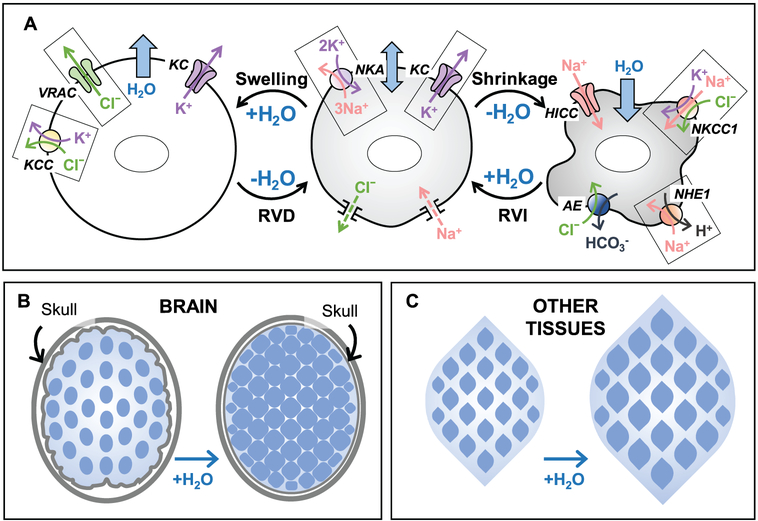
Figure 12.1.
Basic principles of cell volume regulation in mammalian cells and major differences in the osmotic homeostasis within the brain and in other tissues. (A) Ion transport mechanisms responsible for cell volume regulation. Center panel: Under isosmotic conditions, the work of the Na+,K+-ATPase (NKA) and the dominant activity of K+ channels (KC), set the transmembrane ionic gradients and negative membrane potential, as well as compensate for the persistent Na+ uptake via a variety of mechanisms (Na+ leak). Importantly, the negative membrane potential drives out intracellular Cl−, offsetting the presence of the negatively charged impermeant macromolecules and metabolites in the cytosol. Left panel: Hypoosmotic cell swelling triggers activation of the volume-regulated Cl−/anion channel (VRAC) and several K+,Cl− cotransporters (KCC). Cooperative activity of VRAC, KC, and KCC mediates the loss of cytosolic KCl and powers regulatory volume decrease (RVD). Right panel: Cell shrinkage upon exposure to hyperosmotic media stimulates the ubiquitous Na+,K+,Cl− cotransporter 1 (NKCC1) and/or the Na+/H+ exchanger 1 (NHE1). NHE works in cooperation with the volume-insensitive Cl−/HCO3−anion exchangers (AE). In some cell types, cell shrinkage also opens hypertonicity induced non-selective cation channels (HICC). The combined activity of these transporters and channels leads to cytosolic accumulation of NaCl and KCl and mediates regulatory volume increase (RVI). (B and C) The major differences in cell volume control between the brain and other tissues are due to the fixed volume of (extracellular + intracellular) space in the CNS. (B) In the brain, due to spatial limitations imposed by the rigid skull, cell swelling under hypoosmotic conditions or in pathologies occurs at the expense of the interstitial volume and may also compress blood vessels and cause ischemia. Also, due to the restricted ion transport across the blood-brain barrier, there is a “fixed” total pool of extracellular and intracellular ions. Therefore, during cell volume regulation, the electrochemical driving forces for ionic fluxes dissipate very quickly. (C) In contrast to the brain, the majority of peripheral tissues are not restricted in terms of their osmotic expansion or shrinkage, and allow for the relatively rapid exchange of electrolytes between the interstitial space and the blood.

