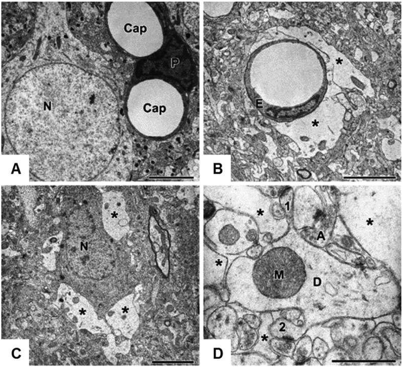
Figure 12.4.
Cell swelling in status epilepticus revealed by electron microscopy (EM). (A) Structural analysis of the control rat neocortex reveals neuronal cell bodies [N], capillaries [Cap], and pericytes [P]. (B and C) Two hours following injection of 4-aminopyridine, EM shows swollen perivascular astrocytic endfeet [asterisks in (B)], which surround an endothelial cell [E]. Swollen astrocytic processes [asterisks in (C)] also border an apparently shrunken pyramidal neuronal body [N]. (D) A higher magnification image in the CA3 layer of the hippocampus displays a swollen dendrite [D] with apparently normal mitochondrion [M], and adjacent electron-transparent swollen astrocytic processes [asterisks]. Additionally labeled are: an axon terminal [A] and dendritic spines [1,2]. The scale bars in the fields (A-C) = 5 μm, and in (D) = 1 μm. Reproduced from P.F. Fabene et al., 2006, with permission.

