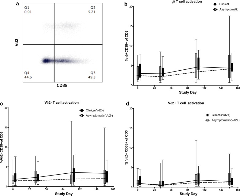
Fig. 6.
Gamma delta T cell activation during the malaria transmission season. a Representative flow cytometry plot of CD38 expression in Vδ2+ and Vδ2− γδ T cells. b Comparison of the total γδTCR + CD38 + T cells. c Vδ2− γδTCR + CD38 + and d Vδ2+ γδ T cells between clinical malaria cases versus asymptomatic infections. All values are expressed as percentage of total CD3 T cells. The data are represented as Box and Whisker plots of medians with ranges. The trend lines indicate the medians for each group

