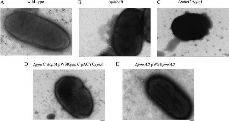
FIG 6.
Transmission electron micrograph of C. rodentium cells producing OMVs. C. rodentium wild-type (A), ΔpmrAB (B), ΔpmrC (ΔeptA) ΔcptA (C) ΔpmrC (ΔeptA) ΔcptA pWSKpmrC/eptA pACYCcptA (D), and ΔpmrAB pWSKpmrAB (E) cells were grown to mid-log phase in N-minimal medium supplemented with 50 μM FeCl2. Culture dilutions in PBS were laid onto carbon-coated copper grids and stained as described in Materials and Methods. Samples were imaged using an accelerating voltage of 75 kV and at a magnification of ×40,000. Images shown are representative of 12 micrographs per strain. Bars, 100 nm.

