Fig. 2.

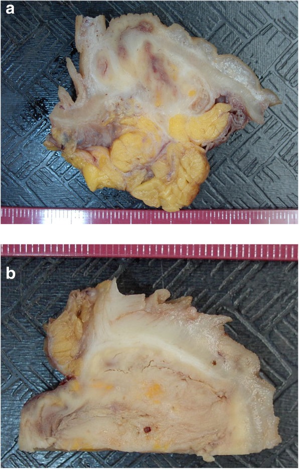
Macroscopy of the resected sigmoid. a and b In both images the mucosa is at top. The underlying bowel wall and mesentary are infiltrated and distorted by malakoplakia infiltrates which are friable and have visible cracking artefact

Macroscopy of the resected sigmoid. a and b In both images the mucosa is at top. The underlying bowel wall and mesentary are infiltrated and distorted by malakoplakia infiltrates which are friable and have visible cracking artefact