
Fig. 3.
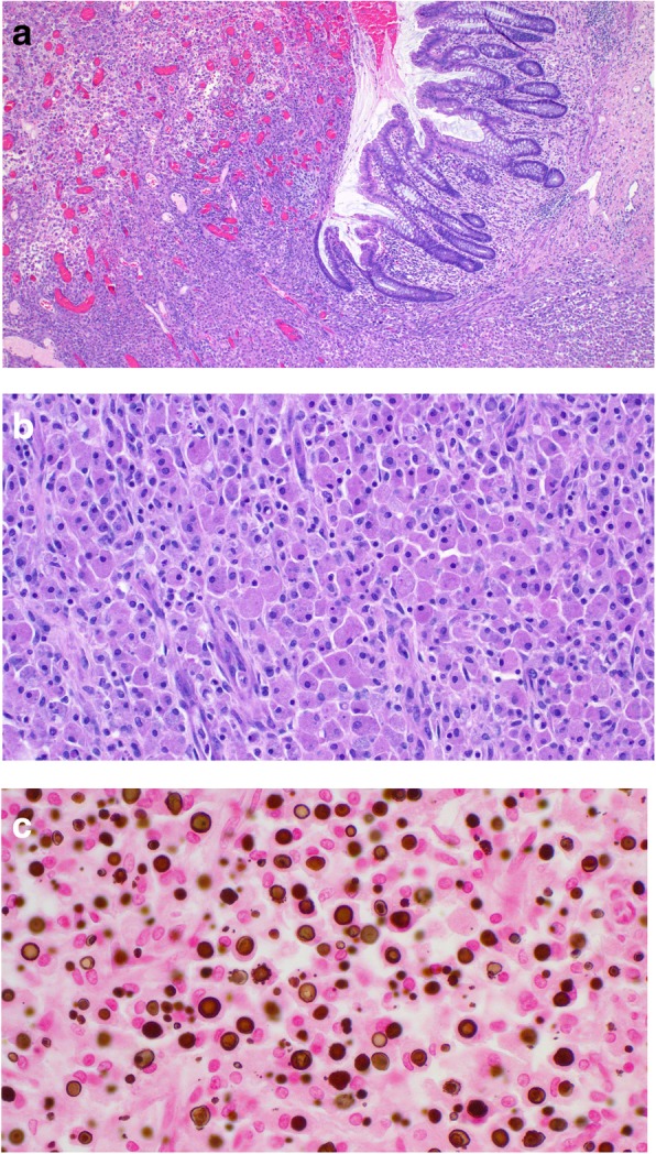
Microscopy of the resected sigmoid. a Low power image showing ulceration of the mucosa due to massive infiltration of all levels of the bowel wall by malakoplakia. b Medium power image of the sheet-like infiltrate of large non-atypical macrophages with abundant granular cytoplasm. c High power image of abundant MG bodies. They are of variable size with a round to ovoid shape. Many have a targetoid appearance