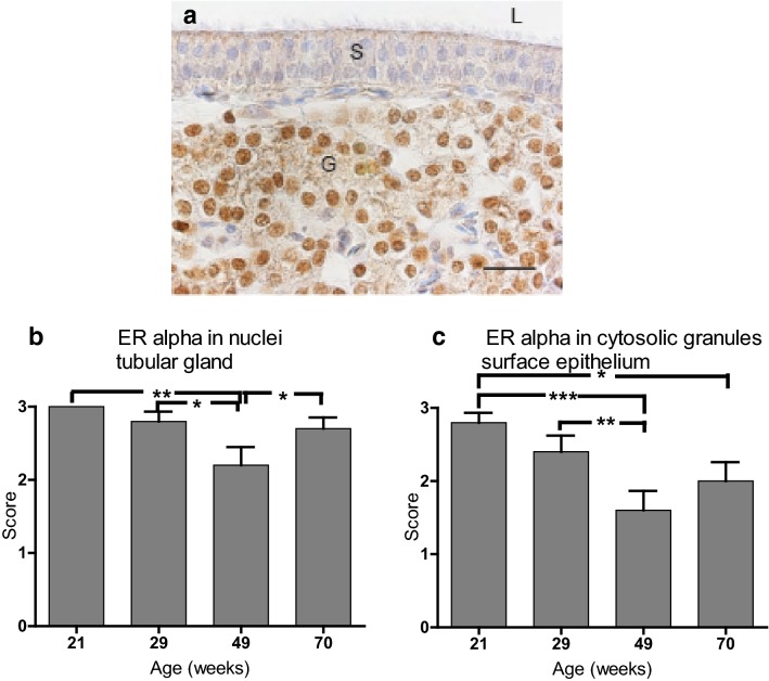
Fig. 6.
Oestrogen receptor α (ERα) immunolocalization in shell gland of Lohmann Selected Leghorn (LSL) and Lohmann Brown (LB) hens at different ages during a production period. a Shell gland mucosal fold of 29-week-old LSL hen showing strong staining of nuclei in tubular gland cells (G), moderate cytosolic granular staining for ERα in surface epithelium (S) and unstained nuclei of both ciliated cells and non-ciliated cells. Lumen (L), bar = 20 µm, weak haematoxylin counterstain. b, c Intensity scoring of oestrogen receptor α (ERα) in shell gland mucosal fold of LB and LSL hens (n = 10) at four different ages during a production period. Score 0: no staining. Score 1: weak staining. Score 2: intermediate staining. Score 3: strong staining. b ERα staining in nuclei of tubular glands. c ERα staining of cytosolic granules in surface epithelial cells. Mean ± standard error. *P < 0.05, **P < 0.01, ***P < 0.001

