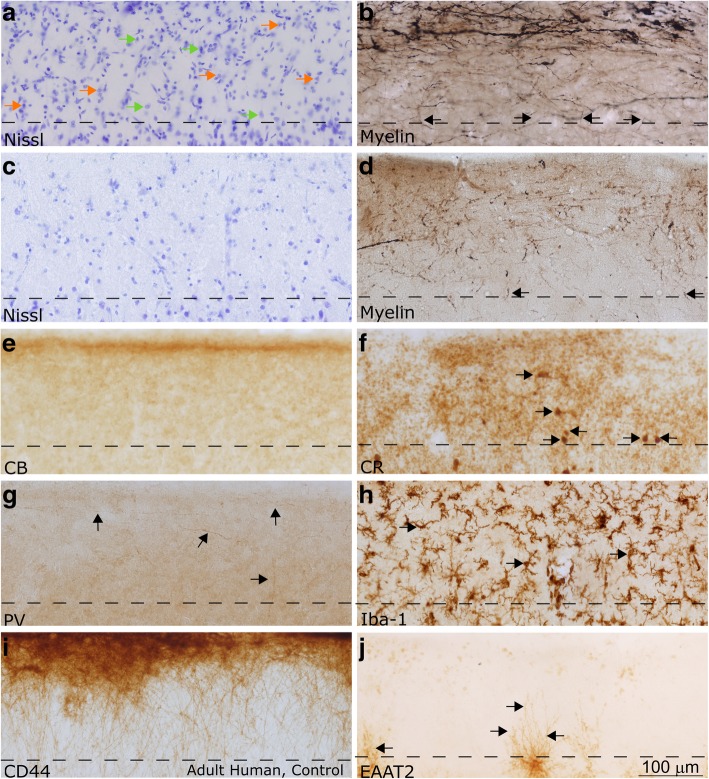
Fig. 7.
Layer 1 in the LPFC and ACC of the adult human. All images in this figure were acquired from neurotypical adult cases (HCD, HCF, HAW, HAY). a-d Nissl (a) and myelin (b) in the LPFC showed stark differences from ACC (c-d). ACC had a thicker layer 1, and reduced density and thickness of myelinated axons in layer 1. Permeating blood vessels and endothelial cells were also visible in the Nissl-labeled section from LPFC layer 1 in (a). Examples of neurons in (a) are marked with orange arrows, glial cells are marked with green arrows. Myelin (Gallyas) stained tissue (b, d) showed a dense plexus of myelinated axons in superficial layer 1, consistent with observations in the non-human primate. While the majority of axons were horizontal, some also had diagonal trajectories, consistent with axons from incoming pathways (black arrows). e-g CB, CR, and PV labeled inhibitory interneuron classes. CB (e) labeled scant processes in layer 1 (labeled with black arrows), while CR (f) labeled a low density of neurons and few processes in layer 1 (labeled with black arrows). PV (g) labeled processes that joined the plexus of axons in superficial layer 1 (labeled with black arrows). h Iba-1 labeled microglia within the cortex, including layer 1 (examples of microglia are labeled with black arrows). Superficial microglia extended processes mostly parallel to the pial surface, while microglia deeper in layer 1 had processes oriented in multiple directions. i CD44 labeled interlaminar astrocytes, which sat on the pial surface and sent processes towards layer 2. These astrocytes are exclusive to higher-order primates. j Astrocytes labeled with excitatory amino acid transporter (EAAT2) were not present in layer 1; however, the processes of labeled astrocytes from layer 2 penetrated into the deep part of layer 1 (labeled with black arrows). Images were acquired such that the top edge of the images underlie the pia. Dotted lines indicate the border with layer 2 in all panels. Calibration bar in (j) applies to all panels

