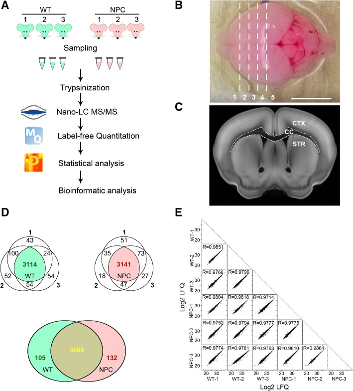
Fig. 1.
Comparison of protein patterns in corpora callosa of wildtype and Npc1 mutant mice at P12. a: The schematic diagram of the workflow for proteomic analysis from 3 biological replicants of wildtype (WT) and Npc1 mice (NPC). b, c: Separated brain was cut according to the dashed lines in the forebrain and the corpus callosum (CC) in the transaction was separated with the cortex (CTX) and the striatum (STR) from sections 2 to 4 in b. Scale bar: 1 cm in b. c was adapted from the Allen Brain Atlas (http://atlas.brain-map.org/). d: The Venn diagram of identified proteins from each sample. Valid proteins from each genotype were illustrated in green for WT samples and in red for NPC samples. The common proteins from both WT and NPC were in yellow. e: The Scatter plots of Log2 LFQ values of identified proteins between samples and the Pearson correlations were calculated (the values of R)

