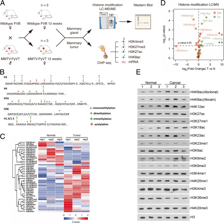
Fig. 1.
Several histone modifications altered in breast cancer development. a Workflow for the whole project, including mice mating, LC-MS/MS screening, Western blot validation, and ChIP-seq analysis. b Overview of quantified histone PTMs in core histone proteins in breast tissues. c Heat map of quantified histone marks found to be regulated in tumor tissue as compared to normal tissue. d Volcano plots representing the fold change (x-axis) and the significance (y-axis) for single histone marks. The statistical difference is calculated using the t test. The two histone marks H3K4me3 and H3K27ac marked with asterisk (*) were only detected in tumor by MS analysis. e Western blot analysis with the indicated histone marks antibodies. Whole tissue lysates were prepared from normal tissue or tumor 3 replicates

