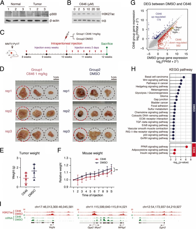
Fig. 7.
p300 inhibitor C646 could suppress breast cancer development in MMTV-PyVT mice. a Western blot analysis with p300. Whole tissue lysates were prepared from normal tissue or tumor three replicates. b Western blot analysis with H3K27ac after adding p300 inhibitor C646 in different concentration (0, 2, 5, 10, 20, 50 μM) to HEK293 cell line. Cell was treated with C646 for 12 h. c Sketch showing the schedule of C646 injection. d Photos of MMTV-PyVT mouse mammary tumors grown 12 weeks. The successive three tumors in a rectangle photo belong to one female MMTV-PyVT mouse, and they were the biggest three tumors in the mouse. All dashed circles represent the same size and the scales locate in the top right corner of each group. The tissues with red circles were subjected for RNA-seq. e Scatter plot showing the tumor weight of the two groups. Summation weight of the biggest three tumors was considered as the tumor weight of each mouse. f Line plot showing the body weight development of mouse in the two groups. **p < 0.001; t test; n = 4. g Scatter plot comparing upregulated genes expression (log2(FPKM + 2−5)) between DMSO group and C646 group. h Matchstick plots showing the top 20 (top) and all three (bottom) KEGG pathway terms of DMSO group and C646 group upregulated genes. i UCSC browser view showing the ChIP-seq density of H3K27ac and RNA-seq signal in both DMSO group and C646 group located in Vegfa, Mrps7, and Egln3 gene locus

