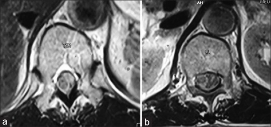
Figure 14.

(a) Magnetic resonance imaging T1-weighted axial image at the level of T12 vertebra showing isointense lesion in the spinal canal arising from the left side pushing the cord toward the right. (b) T1-weighted axial image at the level of L1 vertebra showing the isointense lesion arising from the left side of the spinal cord with mass effect
