Figure 4.

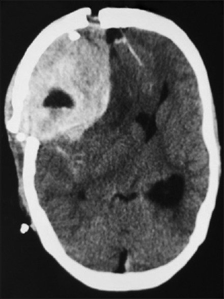
Computed tomography brain plain – showing further increase in the size of extradural hematoma in the left frontal extradural region causing severe mass effect and midline shift

Computed tomography brain plain – showing further increase in the size of extradural hematoma in the left frontal extradural region causing severe mass effect and midline shift