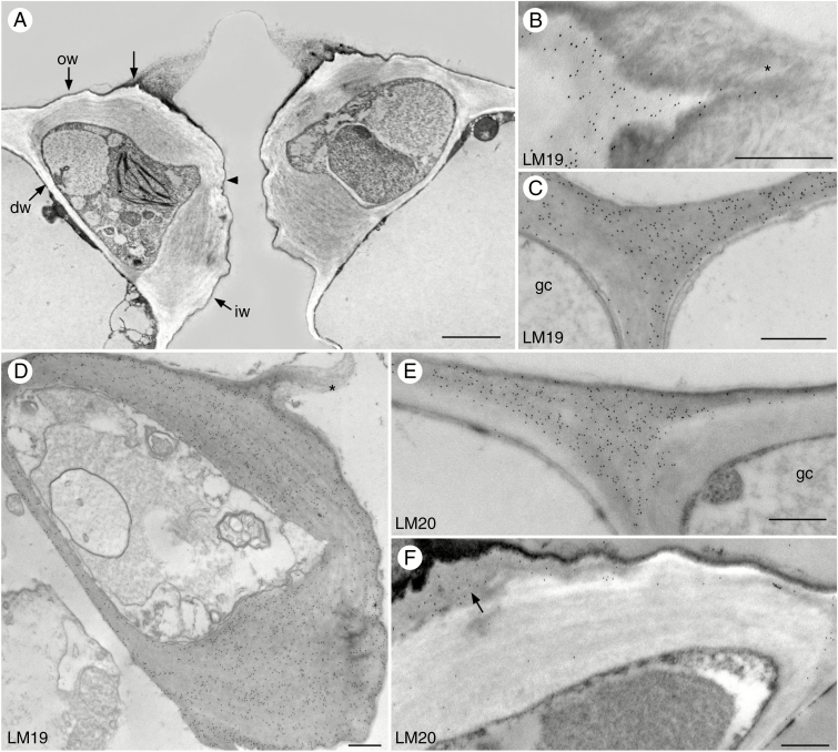
Fig. 1.
Immunolocalization of homogalacturonan pectin epitopes in guard cell walls of Arabidopsis. Black round dots are colloidal gold labels attached to the specific pectin antibody. (A) Cross-section of guard cells showing outer walls (ow), outer ledges (unlabelled arrow), inner walls (iw) and ventral walls which are thinner at midsection (arrowhead). Dorsal walls (dw) are thin and abut epidermal cells. (B) The outer ledge labels with the LM19 antibody and the electron-dense cuticular extension (*) is unlabelled. (C) LM19 antibody labels guard cell (gc) walls and adjacent epidermal cell walls. (D) LM19 label is evenly distributed over the guard cell walls. The outer ledge is extended with cuticle-like material (*). (E) LM20 antibody strongly labels the junction of guard cell (gc) and adjacent epidermal cell wall. (F) LM20 antibody labels the outside wall of the guard cell and outer ledges (arrow), but there is less labelling in the electron-lucent internal thick wall. Scale bars: (A) = 2 µm; (B–F) = 500 nm.

