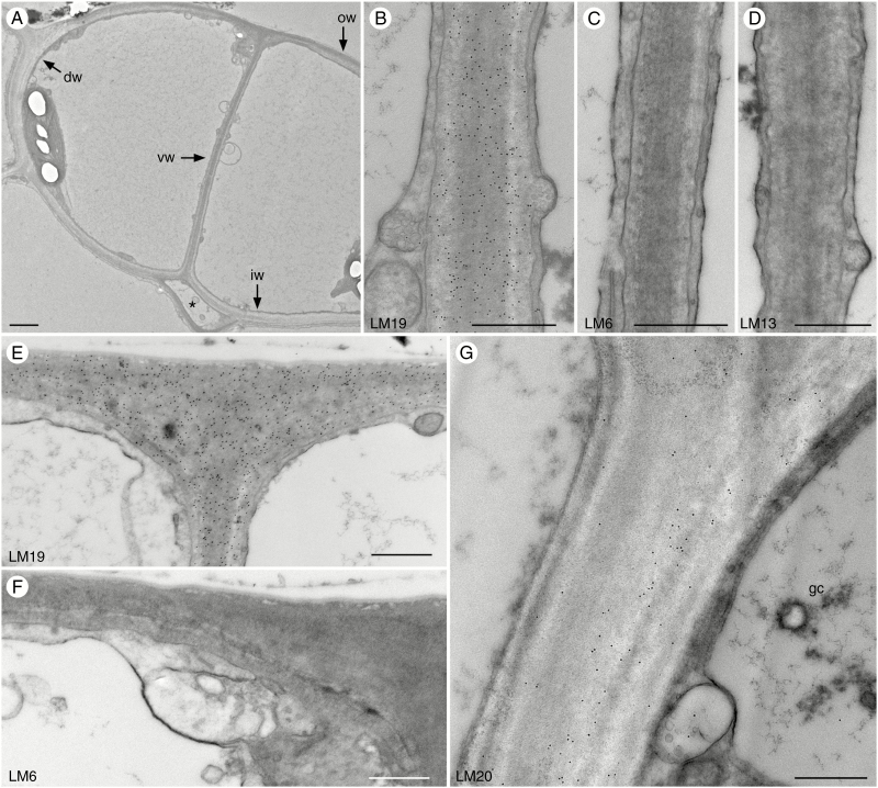
Fig. 3.
Immunolocalization of pectin epitopes in young guard cell walls of Phaeoceros. Black round dots in images are colloidal gold labels attached to the specific pectin antibody. dw, dorsal wall; iw, inner wall; ow, outer wall. (A) Guard cell walls in pre-opened stomata are thin. The substomatal cavity (*) develops before the pore forms along the ventral wall (vw). (B–D) Ventral walls of pre-opened stomata. (B) LM19 antibody labels ventral walls strongly and evenly. (C) No labelling for LM6 antibody was detected. (D) No labelling was detected for LM13 antibody. (E) LM19 antibody strongly recognizes outer walls. (F) No labelling was detected for LM6 antibody in outer walls. (G) LM20 antibody only labels the dorsal wall of guard cells (gc). Scale bars: (A) = 2 µm; (B–G) = 500 nm.

