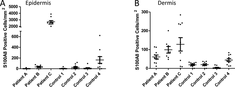
Fig. 2.
Quantification of S100A8 positive cells in the skin of LD patients. S100A8 positive cells were quantified in the epidermis (A) and dermis (B) of skin sections from 3 LD patients and 4 control surgical patients. Results represent cell counts (positive for both DAPI and S100A8) from 8–11 individual images spanning the tissue section of an individual patient. Image numbers: Patient A n=11; patient B n=8; patient C and all four controls n=10. (A) S100A8 staining in the epidermis of patient A is significantly higher than all four controls (p <0.001) and individual patients B and C (p<0.001). (B) S100A8 staining in the dermis of patient C is significantly higher than all four controls (p <0.01); the same is true for patient B and controls 1–3 (p<0.05).

