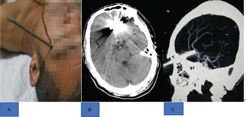
Figure 2.

A, Preoperative CT (computed tomography) scan of the patient's head. Small intraventricular hemorrhage on axial head CT scan showing 1 radiopaque welding electrode penetrating both skull plates and entering the brain parenchyma. B, Welding electrode penetrating the brain from the outer sidewall of the left eye socket. C, CT (computed tomography) sagittal reconstruction, no significant damage to important nerves and vessels was observed.
