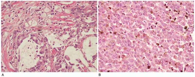
Figure 4.

HE of GC and PMME. (A) The pathology results of the gastric body showed poorly differentiated adenocarcinoma, parts of mucinous adenocarcinoma, and signet ring cells. (B) The esophageal tissues were inflitrated by malignant melanoma (magnification: 200×). HE = hematoxylin–eosin, GC = gastric cancer, PMME = malignant melanoma of the esophagus. PMME = primary malignant melanoma of the esophagus.
