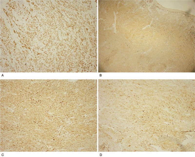
Figure 5.

IHC staining of GC and PMME. (A) Positive staining of Her-2 in GC (magnification: 100×). (B) Positive staining of Melan-A (+) (magnification: 40×), (C) HMB-45 (magnification: 100×), and (D) S-100 (+) (magnification: 100×) in PMME. IHC = immunohistochemistry, GC = gastric cancer, PMME = malignant melanoma of the esophagus. PMME = primary malignant melanoma of the esophagus.
