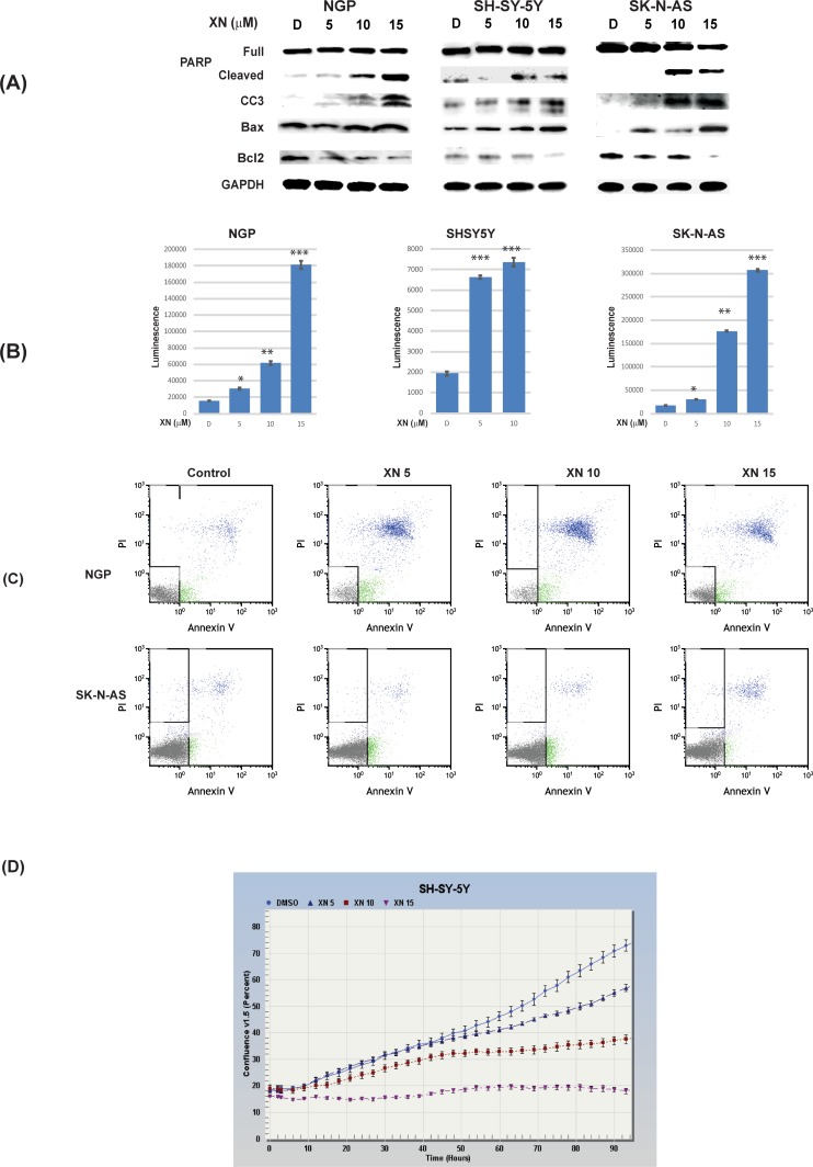
Fig 3. XN induces apoptosis in NGP, SH-SY-5Y, and SK-N-AS cells.
A. Western blot analysis showed an increase in apoptotic markers, cleaved PARP, cleaved caspase 3 (CC3) and Bax after treatment with XN for 72hrs. This was associated with a reduction in anti-apoptotic protein, Bcl-2. B. Caspase-3/-7 activities increased in a dose-dependent fashion in XN-treated NGP, SK-N-AS, and SH-SY-5Y cells. * p<0.05; ** p<0.001; *** p<0.0001. (C). Annexin-V-FITC flow cytometry analysis showed an increase in apoptosis. (D). Live cell image of cell player with YOYO-1 assay by Incucyte also showed induction of apoptosis.

