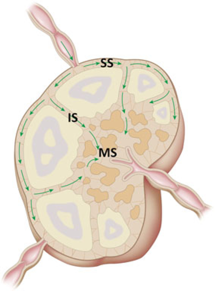
Figure 4.

Schematic illustration of lymph flow observed in this study. Lymph fluids enter the periphery of the lymph node and spread along sub-capsular sinuses before entering into deeper areas, such as the medullary sinuses. SS, subcapsular sinus: IS, intermediate sinus: MS, medullary sinus.
