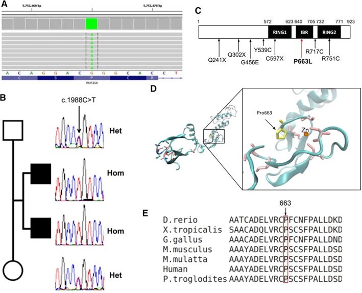
Figure 2.

Molecular and bioinformatic results. (A) Exome result of patient 1 in BAM format, the variant is found in the homozygous state. (B) Sanger results of gene RNF216 for the core family. (C) Relative location of the presently found variant together with other reported missense and nonsense pathogenic variants in the RNF216 gene isoform‐2. (D) Model of zinc rings and IBR domain (modeled using 4KBL structure as template). Cystein residues coordinating the zinc ion are shown in pink; mutated proline in yellow. (E) Multiple alignment of proteins homologous to RNF216 isoform‐2 in different species.
