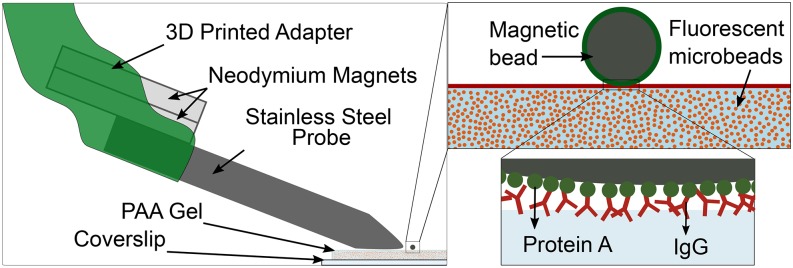
FIG. 1.
Schematic depiction of the magnetic pulling cytometer set-up (left), showing the 3D printed adapter, permanent neodymium magnets, 416 stainless steel probe, and the ‘magnetic bead on gel’ arrangement with a 4.5 μm superparamagnetic bead bound to a polyacrylamide (PAA) gel attached to a glass coverslip. Insets on the right show the magnetic bead coated in protein A (green) atop the PAA gel coated with IgG (red). Fluorescent beads are embedded within the gel to track the force induced displacements in the gel. Protein A binding to the IgG facilitates magnetic bead binding to the PAA gel in the ‘magnetic bead on gel’ arrangement.

