
Figure 2. Loss of the C-terminal domain of DAK leads to altered cell morphology.
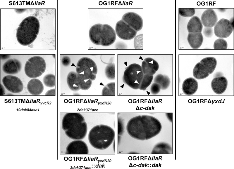
Transmission electron microscopy (TEM) of parent strains (top row), derivative strains (middle row), and complemented strains (bottom row) displaying changes in cell envelope structure. In OG1RFΔliaR septa are positioned at the middle of the cell, with one septal event per cell. In OG1RFΔliaRyxdK202dak371ace, multiple septal events per cell can be seen (white arrows), with loss of the division plane orientation, resulting in distorted cells. Cell envelope at the site of division is thickened, with small burs or protrusions from the cell surface (black arrows). A similar phenotype can be observed in OG1RFΔliaRΔc-dak, suggesting this domain plays an important role in cell division. These changes were not seen when the full length dak gene was complemented back to its chromosomal location. S613TMΔliaRyvcR219dak84asa1 and OG1RFΔyxdJ did not show structural changes. Scale bar for all images, 50 nm; Images are representative of over 25 different fields, wide field images are displayed in supplementary figure 3.