
Figure 3. DAP-R independent of LiaR does not result in phospholipid microdomain redistribution or a reduction in DAP binding.
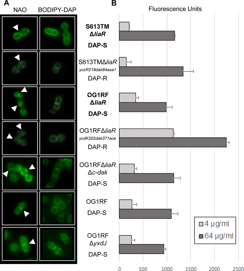
(A) N-nonyl acridine orange (NAO) and BODIPY-DAP staining of E. faecalis strains. NAO is a membrane dye that associates with anionic phospholipids, particularly cardiolipin. In all strains NAO and BODIPY-DAP can be seen localizing to the division septum (white arrows), without any areas of redistribution. (B) Quantitative BODIPY-DAP binding. Comparisons in binding were made between parental stains (bold) and derivatives. Increased DAP binding was seen in OG1RFΔliaRyxdK202dak371ace at both 4 and 64 µg/mL as compared to OG1RFΔliaR. No significant changes were seen between the other strain pairs. Representative results of two independent runs performed on different days.