Figure 4.
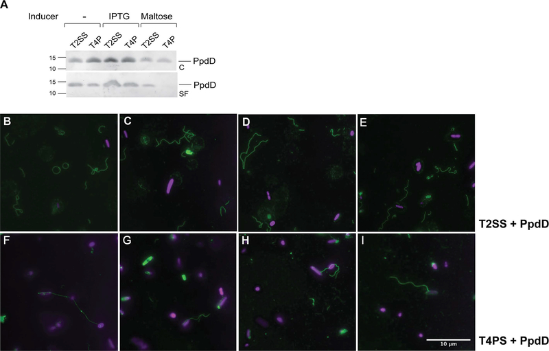
Comparison of PpdD pilus assembly via the T2SS and T4PS. A, E. coli strain BW25113 F’lacIQ containing plasmid pCHAP8565 and pCHAP8184 (T2SS) or pMS41 (T4PS) was grown on LB plates containing AP and Cm at 30°C for 48 hours, without or with indicated inducers. Fractionation and Western blot with antibodies raised against MalE-PpdD fusion protein were performed as described in Experimental procedures. Migration of molecular weight markers is indicated on the left. B-E, Immunofluorescence microscopy analysis of PpdD pili assembled by the T2SS or by the T4PS (F-I). Samples were processed as indicated in Experimental procedures. Pili are labelled in green (GFP) and bacterial nucleoid in magenta (DAPI). Scale bar indicates 10 μM.

