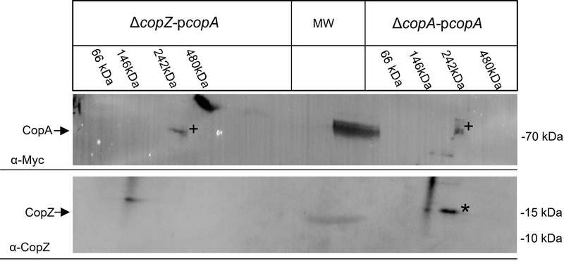
Fig. 7: CopZ forms also a complex with the P1B-type ATPase CopA.
ICMs of ΔcopZ pcopA and ΔcopA pcopA were solubilized, and first separated by BN-PAGE. The BN-PAGE lanes were then cut and subjected to a 2nd dimension SDS-PAGE, followed by immune detection, as described for Fig. 6. CopA and CopZ are indicated by (+) and (*), respectively.

