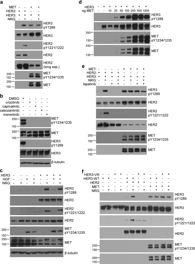
Fig. 1.

Overexpression of MET drives HER3 phosphorylation independently of EGFR and HER2. (a) COS7 cells expressing either MET or HER2 with HER3 were stimulated −/+ NRG (50ng/mL) and assayed for HER3 phosphorylation by western blot. (b) COS7 cells expressing MET and HER3 were treated with DMSO, or MET inhibitor (1μM crizotinib, 100nM capmatinib, 100nM cabozantinib, or 1μM merestinib) for 6 hours and assayed for MET and HER3 phosphorylation by western blot. (c) COS7 cells transfected with onlyHER3 were stimulated with either NRG (50ng/mL), HGF (50ng/mL), or both, and assayed for HER3 phosphorylation and endogenous levels of MET and HER2 by western blot. (d) COS7 cells were transiently transfected with HER3 and increasing amounts of MET and assayed for HER3 phosphorylation and MET expression by western blot. (e) COS7 cells expressing either HER3 + HER2 or HER3 + MET were treated −/+ NRG (50ng/mL) and −/+ 3μM lapatinib and assayed for HER3 phosphorylation by western blot. (f) COS7 cells expressing either wild-type (WT) or V926R mutant (VR) HER3 together with HER2 or MET were stimulated −/+ NRG (50ng/mL) and assayed for HER3 phosphorylation.
