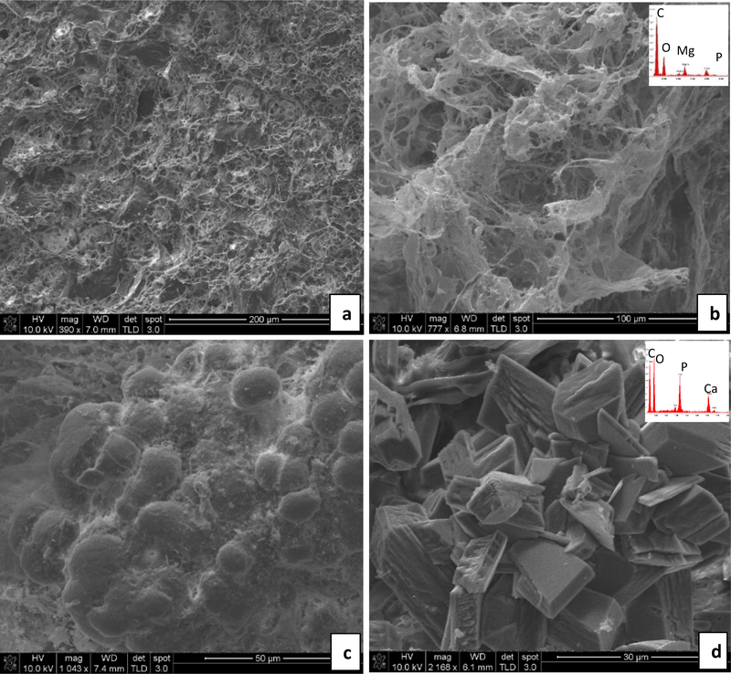
Fig. 4.
Representative SEM images and energy dispersive spectroscopy (EDS) spectra (insets) obtained from stones overgrown on the implants– Regular water group. (a & b) DNM group: In (a), the tissue matrix contains a small amount of magnesium phosphate mineral without visibly obvious crystals, even though the EDS spectrum (b inset) detects these elements. (c & d) BRP-C group: In (c), a calcium phosphate spherulitic coating is comprised of fine crystals, probably those of the original BRP but with an accumulation of some organic debris. In (d), very large calcium phosphate crystals, unlike the morphology of the crystals in the original BRP, have formed. Scale bars are as follows: (a) 200 μm, (b) 100 μm, (c) 50 μm, (d) 30 μm

