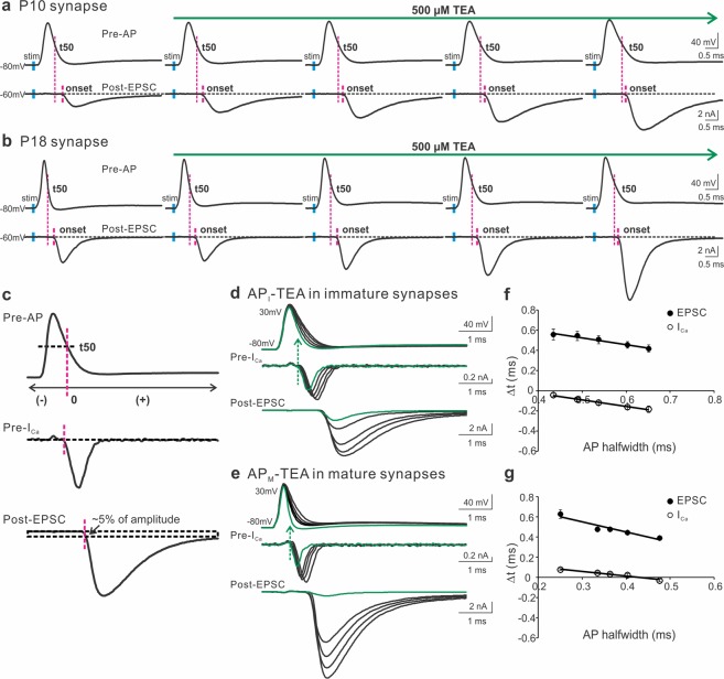
Figure 1.
The contribution of presynaptic K+ channels to the onset of ICa and EPSC. (a,b) A representative AP (top panels) and EPSC (bottom panels) recorded from pre- and postsynaptic compartments of the calyx of Held synapse in response to axonal stimulation (blue bars) applied to a brain slice taken from a mouse at P10 (a) or P18 (b). TEA (500 μM) in the external solution containing 2 mM Ca2+ was perfused to gradually block K+ channels. The magenta lines indicated the onset of EPSC relative to the half decay time (t50) of APs. (c) t50 was defined as the repolarization time at the half-maximal amplitude of an AP and was set as time zero. Before or after t50 had negative or positive values. The onset of ICa was marked as the beginning of the inward current below the baseline. The onset of EPSC was determined by the rise within 5% of their amplitude. (d,e) Paired recordings of ICa and EPSC from immature (d) and mature (e) synapses evoked by the AP templates previously recorded from the P10 (a) and P18 (b) synapses, respectively. The currents produced by real APs before TEA exposure were highlighted in green. The extracellular solution included 1 mM Ca2+ to improve the quality of recordings. (f,g) Summary plots of the onset timing of ICa (empty circles) or EPSC (black circles) relative to t50 against the AP halfwidth for immature (n = 11, f) and mature synapses (n = 6, g). Solid lines were linear regressions of the data.

