Figure 2.
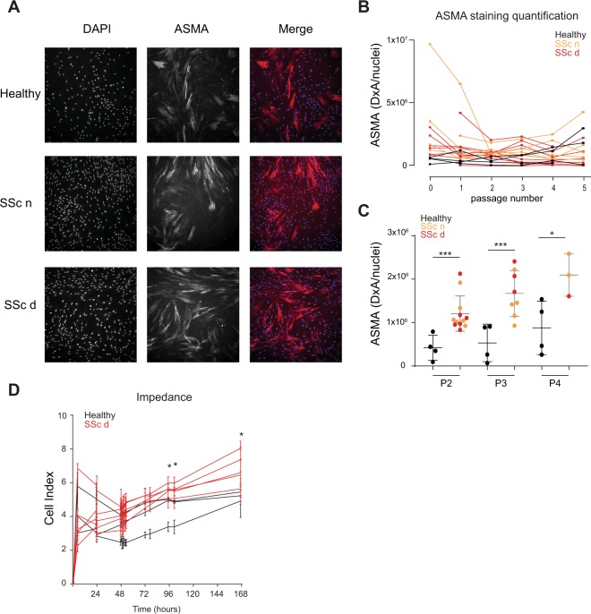
High ASMA expression and abnormal cell impedance are detected in primary SSc dermal fibroblasts up to four in vitro culture passages. Primary dermal fibroblasts from SSc donors and healthy donors were immuno-stained to measure ASMA expression and stained with DAPI to detect nuclei. (A) Representative images of the ASMA and DAPI stainings from dermal fibroblasts isolated from a healthy donor and an SSc donor. The SSc dermal fibroblasts were isolated from a clinically affected area (SSc d) and non-affected area (SSc n). (B) Image quantification results of ASMA staining (Density X Area corresponding to the intensity of the staining multiplied by the area) divided by the number of nuclei stained with DAPI of SSc dermal fibroblasts (orange and red) or healthy dermal fibroblasts (black) at different cell culture passages. (C) Differences in ASMA staining (Density X area) divided by the number of nuclei between SSc dermal fibroblasts and healthy dermal fibroblasts at different cell culture passages. (D) Cell index from SSc dermal fibroblasts and healthy dermal fibroblasts after four in vitro cell passage. Statistical significances were assessed using two-tailed Student’s t-tests with *p-value < 0.05, ***p-value < 0.001.

