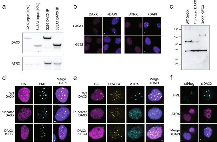
Figure 2.
Loss of C-terminal DAXX sequence drives mislocalization of DAXX-KIFC3 and ATRX. (a) Western blot of DAXX immunoprecipitation. DAXX-KIFC3 was competent to bind ATRX in G292, similar to wild-type DAXX in SJSA1 cells. All cropped images are from the same gel, which was stripped and reprobed. Complete images in Supplementary Fig. S2a. (b) Immunofluorescence imaging showing localization of DAXX and ATRX in ALT + and TERT + osteosarcoma cell lines. In SJSA1 osteosarcoma cells wild-type DAXX and ATRX were localized to nuclear punctae (arrowheads), but in G292 the proteins were observed to be diffusely localized throughout the nucleus. Scale bars represent 2 µm. (c) Western blot showing expression of HA-tagged wild-type DAXX, truncated DAXX and DAXX-KIFC3 in G292. Expected MW were 83 kDa for the HA-tagged truncated DAXX and 129 kDa for HA-tagged DAXX-KIFC3. The band at 70 kDa was non-specific. Blank lanes have been cropped. (d) Immunofluorescence imaging of transiently expressed WT DAXX, truncated DAXX and DAXX-KIFC3 in G292 cells. Exogenous WT DAXX was enriched at PML bodies unlike either truncated DAXX or exogenous DAXX-KIFC3. Scale bars represent 2 µm. (e) Immunofluorescence imaging showing localization of ATRX with transiently expressed WT DAXX, truncated DAXX and DAXX-KIFC3 in G292 cells. ATRX forms nuclear foci colocalizing with wild-type DAXX but remains diffuse in cells expressing exogenous truncated DAXX or the DAXX-KIFC3 fusion. Scale bars represent 2 µm. (f) Immunofluorescence imaging showing localization of ATRX in the absence of DAXX-KIFC3. Knock-down of DAXX-KIFC3 (Supplementary Fig S2b) did not restore ATRX localization to PML bodies. Scale bars represent 2 µm.

