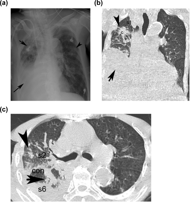
Figure 1.
Presentation of an 82-year-male of [aPTB] with acute lung injury presenting as fever (score = 0), dyspnea (score = 0) in HRCT and [CXR + Hypoalbuminemia] model; Hypoalbuminemia (score = 1) in [CXR + Hypoalbuminemia] model. The CXR shows right (right upper black arrowhead)/left (left upper black arrow) upper lung field patch/nodules, (score = 1) and right lower lung field consolidation with pleural effusion (right lower black arrow) (score = 0) (a). The coronal section of HRCT shows clusters of mass of s1 of right upper lobe (right upper black arrowhead) (score = 2), pleural effusion with consolidation (right lower black arrow) of right lower lobe (b); The transverse section of HRCT shows clusters nodules in s2 (score = 2) of the right upper lobe (right black arrow) and consolidation in s6 of the right lower lobe (right black arrowhead) (score = 1) (c). The total score in [CXR (score1) + Hypoalbuminemia (score1)] model is 2; total score in the HRCT model is 3 [clusters of mass/nodules in s1/s2 of right upper lobe (score2) + consolidation in s6 of right lower lobe (score1)]. c = cluster nodules/mass; cav = cavitation; s1 = apical segment; s2 = posterior segment right upper lobe; s1 + s2 = apico-posterior segment left upper lobe; s6 = superior segment of right or left lower lobe.

