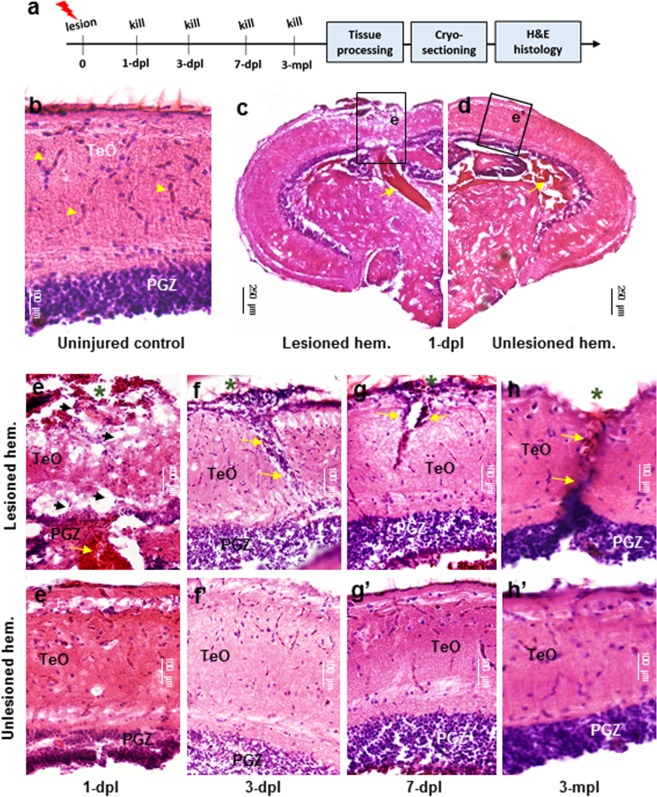
Figure 2.
Histological response following tectal lesion. (a) Experimental design for hematoxylin and eosin (H&E) staining following lesion. (b) Control tectum showing well organized PGZ and highly vascularized superficial TeO layers (yellow arrowheads). (c,d) Overview of the tectal response to stab lesion in the lesioned (c) and unlesioned (d) hemispheres at 1-dpl. At low magnification most conspicuous is the blood pooling immediately below the lesion site and the highly disrupted TeO and PGZ compared to the unlesioned hemisphere. Higher magnification views are demarcated by black boxes for lesioned (e) and unlesioned (e’) hemispheres. (e-e’) Tissue 1-dpl in the lesioned hemisphere (e) showed extensive damage across tectal layers (TeO + PGZ) following cannula insertion, the presence of vacuoles at the lesion site (black arrowheads), and extensive blood pooling underlying the fragmented PGZ (yellow arrows). Despite a slight disruption in the PGZ, the unlesioned hemisphere (e’) closely resembles uninjured control tissue (b). (f-f’) Tissue at 3-dpl in the lesioned hemisphere (f) was characterized by a reduction in the size of vacuoles and oedema, along with a pronounced increase in the number of cell bodies within the lesion canal (yellow arrows). However, already at 3-dpl the PGZ begins to resembled the unlesioned hemisphere. The unlesioned hemisphere (f’) appeared similar to 1-dpl (e’). (g-g’) Tissue at 7-dpl in the lesioned hemisphere (g) demonstrated a reduction in the number of cell bodies from within the lesioned canal, but showed that both sides of the canal have yet to re-joined together (yellow arrows). No change in the unlesioned hemisphere from 3-dpl was noted (g’). (h-h’) While complete tissue recovery was not achieved by 3-mpl in the lesioned hemisphere (h), tissue had re-joined together and both the superficial TeO layers and PGZ closely resembled control conditions. At 3-mpl, the unlesioned hemisphere was indistinguishable from the uninjured control (b). Cross-sectional views with dorsal oriented up are shown for all histological images. In (e–h), green asterisks denote the site of lesion. dpl, days post lesion; mpl, months post lesion.

