Figure 4.
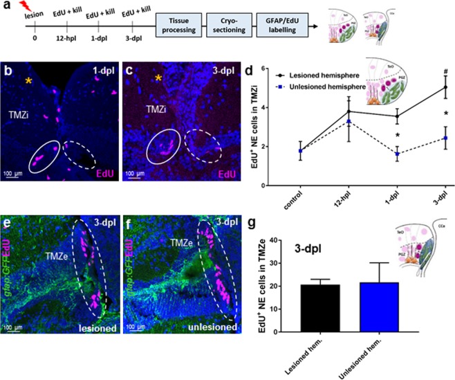
Cell proliferation post-lesion in the TMZi (stem cell zone 2) and TMZe (stem cell zone 3). (a) Experimental design to investigate EdU proliferation by NE progenitors of the TMZi and TMZe. (b,c) Examples of the EdU+ population size of NE amplifying progenitors (pink EdU) in the TMZi observed in the lesioned (yellow asterisk; solid white circle) and unlesioned (dashed white circle) hemispheres at 1-dpl (b), and 3-dpl (c). (d) Number of EdU+ NE cells in the TMZi of lesioned hemispheres at times post-injury compared with control (uninjured animal) levels (one-way ANOVA, F (3, 18) = 7.686, p = 0.0016; Tukey’s multiple comparisons test: control vs 3-dpl, p = 0.005; significance denoted by#), and between lesioned and unlesioned hemispheres at each time point (unpaired t-test, two-tailed: 1-dpl lesioned vs 1-dpl unlesioned, p = 0.0061; 3-dpl lesioned vs 3-dpl unlesioned, p = 0.0096; significance denoted by *). (e,f) Examples of EdU+ labelling (pink) of NE cells in the TMZe of the posterior tectum (white dashed circles) at 3-dpl between the lesioned and unlesioned hemispheres. (g) Quantification of the number of EdU+ cells between the lesioned and unlesioned hemispheres in the TMZe (unpaired t-test, two-tailed: p = 0.9211). Experimental replicates were combined for all statistical analyses. All data presented are mean ± S.E.M. Significance was accepted at p < 0.05 and is denoted by an asterisk unless stated otherwise. In panels b,c,e,f DAPI nuclear counterstaining (blue) was performed. In all cross-sectional images dorsal is oriented up. TMZi, internal tectal marginal zone; TMZe, external tectal marginal zone; hpl, hours post lesion; dpl, days post lesion.

