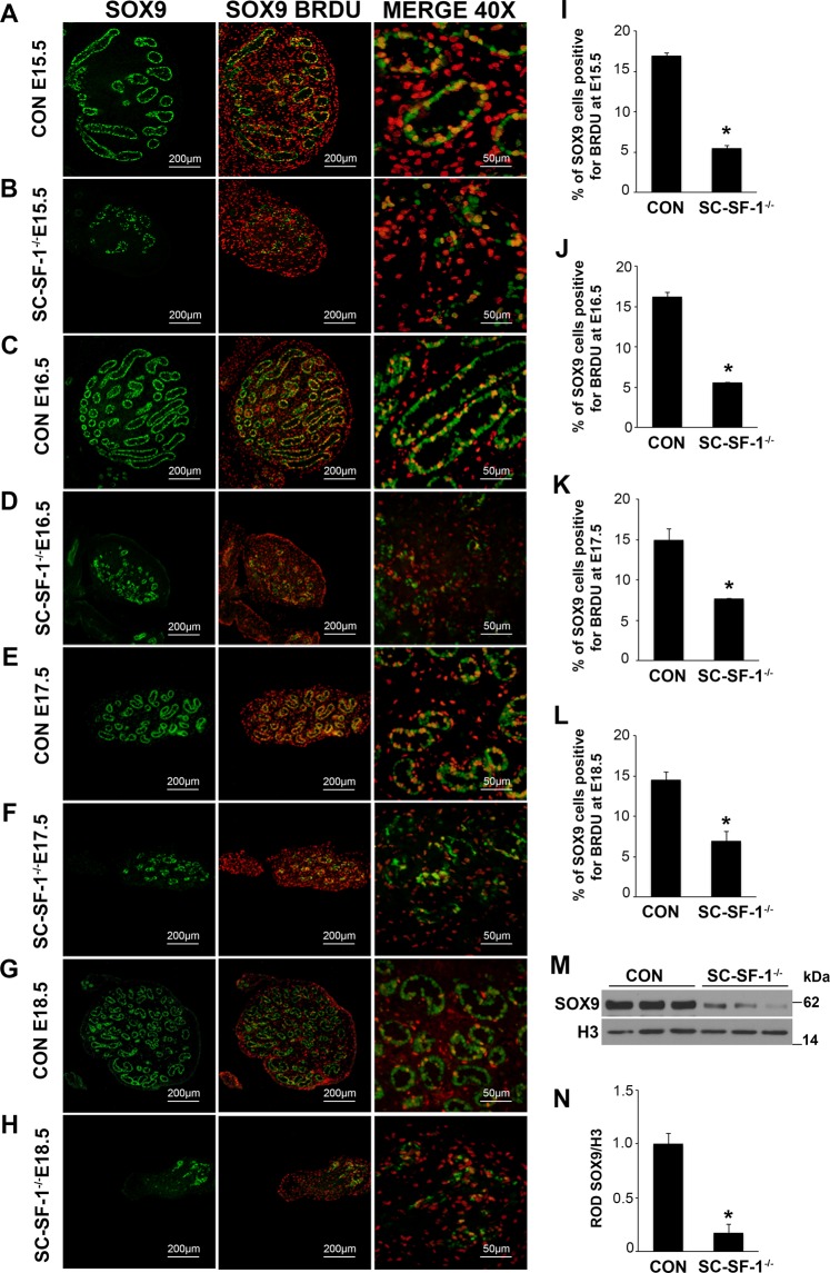
Figure 1.
Reduced expression of SOX9 and loss of proliferation of Sertoli cells in SC-SF-1−/− mice. Immunostaining of SOX9 and BrdU in developing testes of control and SC-SF-1−/− mice at E15.5 (A,B), E16.5 (C,D), E17.5 (E,F) and E18.5 (G,H). SC-SF-1−/− testes demonstrated a marked decline in SOX9 positive cells (B,D,F,H) compared to control testes (scale bar = 200 μm). Pregnant mice were injected with BrdU (50 mg/kg b.wt.) 4 h prior to sacrifice. Reduced BrdU incorporation was observed in SC-SF-1−/− testes (B,D,F,H) compared to controls (A,C,E,G). Proliferating Sertoli cells in the seminiferous tubules were identified with SOX9 and BrdU double positive staining (orange; merge 40X). Reduced proliferating Sertoli cells were observed in SC-SF-1−/− testes through E15.5 to E18.5 (I–L) compared to their respective controls. (M) SOX9 protein levels were analyzed by western blotting in whole tissue extracts of control and SC-SF-1−/− mice at E15.5. Histone H3 served as a protein loading control. (N) Quantification of western blots demonstrated marked decline in SOX9 expression in SC-SF-1−/− testes compared to control testes. *p < 0.001 by two-tailed Student’s unpaired t test. CON = control.

