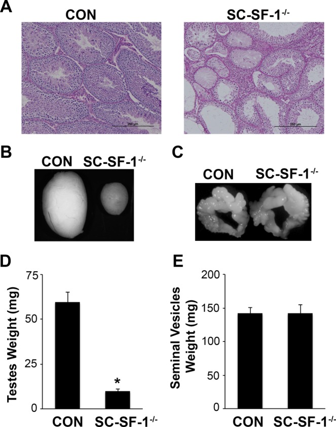
Figure 7.

NR5A1 depletion in Sertoli cells impeded testes development. (A) Histological analysis of testis sections stained with Periodic Acid-Schiff in control and SC-SF-1−/− mice at 6 weeks of age (scale bar = 250 μm). PAS stain showing abnormal phenotype of the seminiferous tubules including vacuolization and visibly large areas of interstitial cells in SC-SF-1−/− testis. Majority of these seminiferous tubules were devoid of germ cells, forming a Sertoli-cell-only phenotype. Stereomicroscopy images of testes (B) and seminal vesicle (C) from control and SC-SF-1−/− mice at 6 weeks. Average weight of the testes (D) and seminal vesicles (E) of control and SC-SF-1−/− mice. The testes were smaller in SC-SF-1−/− mice, while seminal vesicles were similar in size compared to those of control mice. Values are expressed as the mean ± standard error of the mean (SEM) from 5 mice. *p < 0.001 by two-tailed Student’s unpaired t test. CON = control.
