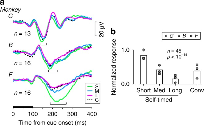
Fig. 2.

Contextual modulation of visually evoked potentials. a Time courses of striatal LFPs aligned with the cue onset in the contralateral visual field. Colored traces indicate the self-timed trials with different interval conditions. Black dashed traces indicate the conventional MS trials. The horizontal black bar denotes the timing of cue presentation. Brackets indicate the ranges of maximal response timing. b Comparison of the magnitude of visually evoked response. Each bar summarizes normalized response obtained from 45 sites. Different symbols plot the means of different monkeys. A one-way repeated measures ANOVA revealed a statistically significant difference across the interval conditions (F2,88 = 50.4, p < 10−14). Conv, conventional memory-guided saccade (MS) task
